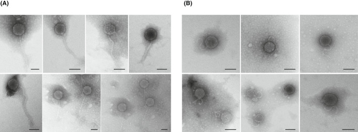
Fig. 1.

Transmission electron micrographs of phages purified from a pool of 10 chicken liver samples from group 2 (sterile inner tissue). Two morphologies were observed: phages with icosahedric capsids and a long non‐contractile tail (A) or a short and curly non‐contractile tail (B). Bar 50 nm.
