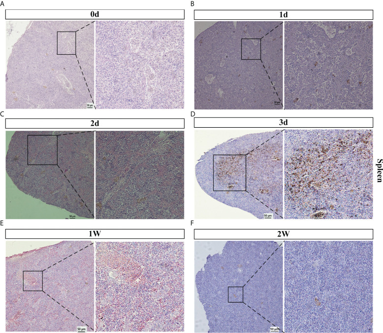
Figure 2.
Spleen histopathology analyses of V. Parahaemolyticus artificially infected with E. coioides. (A–F) The spleen slice of E. coioides artificially infected with V. parahaemolyticus for 0 d (Control), 1 d, 2 d, 3 d, 1 w, 2 w. Representative images from at least three biological replicates of each time point of two groups.

