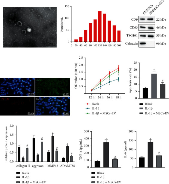
Figure 1.

MSCs-EVs suppresses IL-1β-induced chondrocyte inflammation and ECM degradation in OA. (a) The morphology of EVs observed under the TEM (scale bar = 100 nm). (b) The distribution of EVs analyzed by NTA. (c) Protein levels of CD9, CD63, TSG101, and Calnexin measured by immunoblotting. (d) The uptake of EVs by chondrocytes observed under the microscope (scale bar = 25 μm). Chondrocytes were treated with IL-1β and/or MSCs-EVs. (e) Cell proliferation assessed by CCK8 assay. (f) Apoptosis assessed by flow cytometry. (g) Protein levels of collagen II, aggrecan, MMP13, and ADAMTS5 in chondrocytes measured by immunoblotting. (h) Levels of IL-6 and TNF-α in chondrocytes measured by ELISA. ∗p < 0.05 vs. BMMSCs or chondrocytes without treatment, #p < 0.05 vs. chondrocytes treated with IL-1β. Data are shown as the mean ± standard deviation of three technical replicates. Data between two groups were compared by independent t-test. Data comparisons among multiple groups were analyzed by the one-way ANOVA with Tukey's post hoc test, and comparison of data at different time points was analyzed using repeated measurement ANOVA with Tukey's post hoc test.
