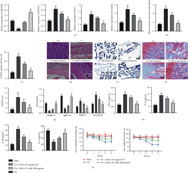
Figure 8.

MSCs-EVs-encapsulated miR-3960 relieves chondrocyte inflammation and ECM degradation in OA mice. OA mouse models were established and injected with MSCs-EVs-miR-3960 agomir (n = 6). (a) miR-3960 expression and mRNA levels of PHLDA2, SDC1, and β-catenin in chondrocytes of OA mice determined by RT-qPCR. (b) Protein levels of PHLDA2, SDC1, and β-catenin in chondrocytes of OA mice determined by immunoblotting. (c) Apoptosis of chondrocytes in OA mice detected by TUNEL assay (200 ×). (d) Articular cartilage injury of OA mice assessed by HE staining (scale bar = 25 μm). (e) aggrecan content of cartilage tissues in OA mice detected by alcian blue staining (scale bar = 50 μm). (f) ECM degradation of cartilage tissues in OA mice assessed by Safranin O-Fast Green staining (scale bar = 100 μm) and OARSI score. (g) Protein levels of collagen II, aggrecan, MMP13, and ADAMTS5 in chondrocytes of OA mice measured by immunohistochemistry. (h) Levels of IL-6 and TNF-α in the serum of OA mice measured by ELISA. (i) PWT and PWL of mice. ∗p < 0.05 vs. sham-operated mice, #p < 0.05 vs. OA mice injected with MSCs-EVs-agomir-NC. Data are shown as the mean ± standard deviation of three technical replicates. Data between two groups were compared by independent t-test. Data comparisons among multiple groups were analyzed by the one-way ANOVA with Tukey's post hoc test.
