
Figure 1. Combined prenatal stressors induce maternal immune activation.
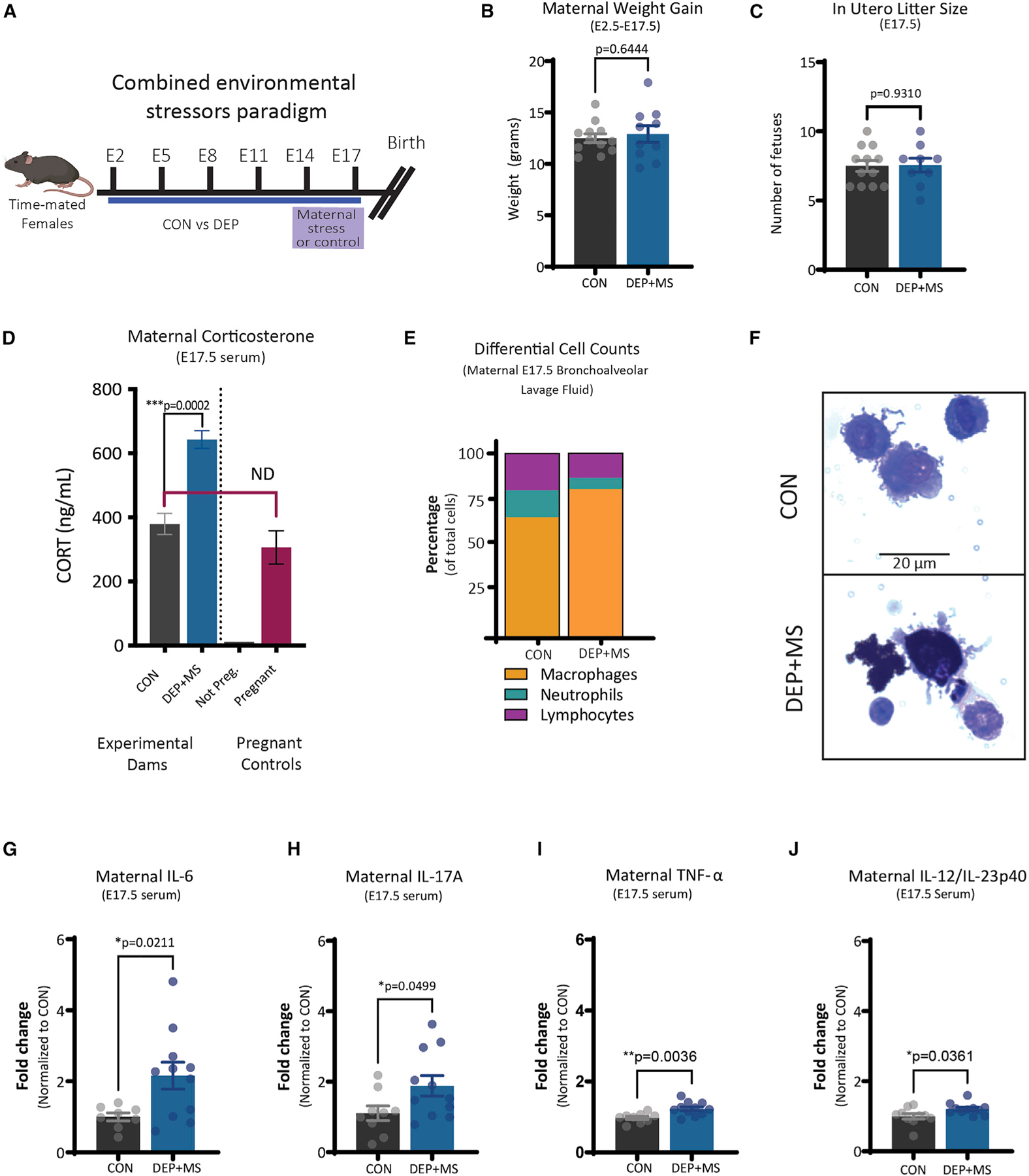
(A) Combined environmental stress paradigm.
(B) Pregnancy weight gain from E2.5 to E17.5 (n = 10–12 mice/condition, unpaired t test).
(C) In utero litter size at E17.5 (n = 10–12 mice/condition, unpaired t test).
(D) Serum concentrations of CORT at E17.5 (left, n = 5–6 mice/condition), and compared with non-pregnant and pregnant WT controls (right, n = 1–3 mice, unpaired t test; ND, not different).
(E) Differential cell count of BALF cells (n = 5 mice/condition).
(F) Representative image of CON versus DEP + MS alveolar macrophages.
(G–J) Serum concentrations of cytokines at E17.5 (n = 10–12 mice/condition, unpaired t test). Means ± SEM.