Fig. 5.
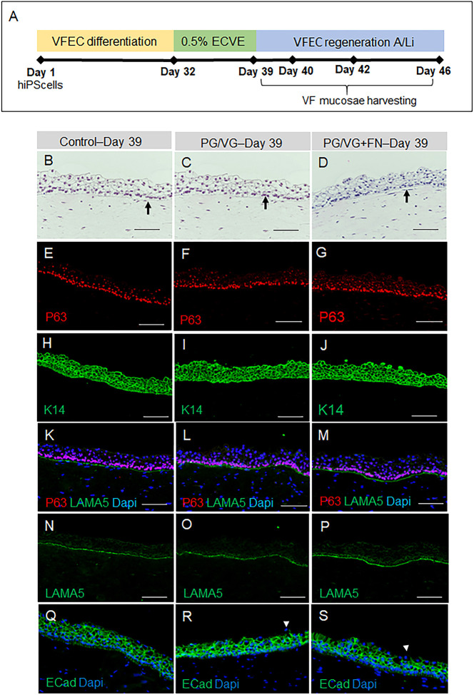
VF epithelial recovery and morphology of hiPSC-derived VF mucosae exposed to 0.5% ECVE at day 39. (A) Schematic illustration of the experimental design for assessment of VF epithelial recovery. HiPSCs were first differentiated into VF epithelial cells (VFECs) for 32 days and then exposed to 0.5% ECVE for 1 week. At day 39 (0 days post exposure), ECVE was withdrawn from the experimental system and VF mucosae were allowed to regenerate for 1 day (day 40), 3 days (day 42) and 7 days (day 46) in regular FAD medium at the A/Li. At selected time points, VF mucosae were harvested and analyzed. (B-D) Morphology of VF mucosae in the control group (B) and 0.5% ECVE-exposed groups (C,D) showing stratified squamous VF epithelia. Black arrows denote the basal cellular compartment. (E-G) Anti-P63 staining (red) in control (E) and 0.5% ECVE-exposed VF mucosae (F,G). (H-J) Anti-cytokeratin 14 staining (green) in control (H) and 0.5% ECVE-exposed VF mucosae (I,J). (K-M) Anti-laminin subunit α-5 (green) co-stained with anti-P63 (red) in control (K) and 0.5% ECVE-treated VF mucosae (L,M). (N-P) Anti-laminin subunit α-5 as a single green channel in control (N) and 0.5% ECVE-exposed VF mucosae (O,P). (Q-S) Anti-E-cadherin staining (in green) in control (Q) and 0.5% ECVE-exposed VF mucosae (R,S). White arrowheads in panels R and S point to apical cells with decreased E-cadherin expression. The histology datasets were performed in three biological and two technical replicates (n=3) and were repeated twice in the laboratory by two investigators. Scale bars: 100 µm.

