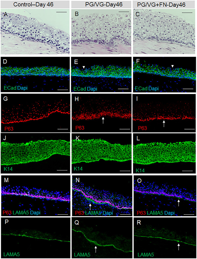
Fig. 6.
HiPSC-derived VF mucosae exposed to 0.5% ECVE at day 46. (A-C) Morphology of VF mucosae in control group (A) and 0.5% ECVE-exposed groups (B,C) at day 46 (7 days post exposure) showing thickened stratified squamous VF epithelia and tightly packed cells in the basal cellular compartment, particularly in the PG/VG group (B). (D-F) Anti-E-cadherin staining (in green) in control (D) and 0.5% ECVE-exposed VF mucosae (E,F). White arrowheads in panels E and F point to apical cells with decreased expression of E-cadherin. (G-I) Anti-P63 staining (red) in control (G) and 0.5% ECVE-exposed VF mucosae (H,I). White arrows in panels H and I denote tightly packed P63+ cells indicating epithelial hyperplasia. (J-L) Anti-cytokeratin 14 staining (green) in control (J) and 0.5% ECVE-exposed VF mucosae (K,L). (M-O) Anti-laminin subunit α-5 (green) co-stained with anti-P63 (red) in control (M) and 0.5% ECVE-treated VF mucosae (N,O). (P-R) Anti-laminin subunit α-5 as a single green channel in control (P) and 0.5% ECVE-exposed VF mucosae (Q,R). White arrows in panels N,O,Q,R denote local thickening of the basement membrane. The histology datasets were performed in three biological and two technical replicates (n=3) and were repeated twice in the laboratory by two investigators. Scale bars: 100 µm.

