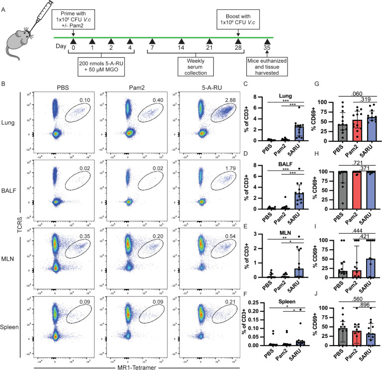
Figure 1.
MAIT cells expand and persist in lungs and bronchoalveolar lavage fluid (BALF) following intranasal 5-A-RU treatment. (A) Live intranasal MAIT ligand plus V. cholerae O1 Inaba vaccination model timeline. (B) Representative FACS plots and (C-F) frequency as a percentage of CD3+ cells of MAIT cells (TCRβ+MR1-Tetramer+) from (C) lung, (D) BALF, (E) MLN, and (F) spleen gated on Live CD3+ CD19- CD44+ cells. (G-J) Frequency of MAIT cells expressing CD69 in (G) lung, (H) BALF, (I) MLN, and (J) spleen. Data are represented as Median with IQR from 3 independent experiments. n=11-12 mice per group. *P< 0.05, **P > 0.01, ***P > 0.001, ***P > 0.0001 by 2-tailed Mann-Whitney U test.

