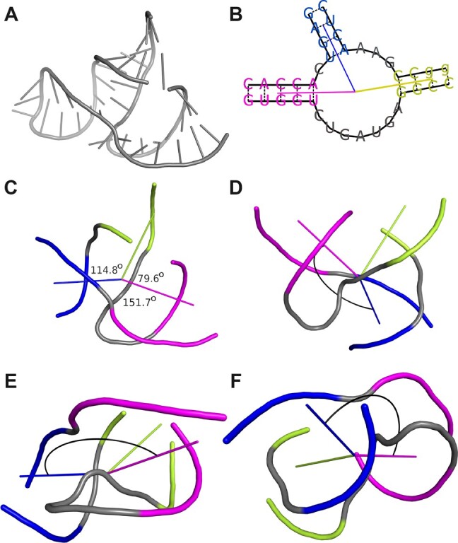
Fig. 2.

(A) The 3D structure of hammerhead ribozyme (PDB ID: 1NYI; Dunham et al., 2003). (B) The 2D diagram of the three-way junction identified in this structure with the directional vectors plotted. (C) The 3D model of thus junction with planar angle values displayed. Euler angles between the blue and the magenta helix shown from the perspective of the (D) X, (E) Y, and (F) Z axes of the coordinate system, respectively
