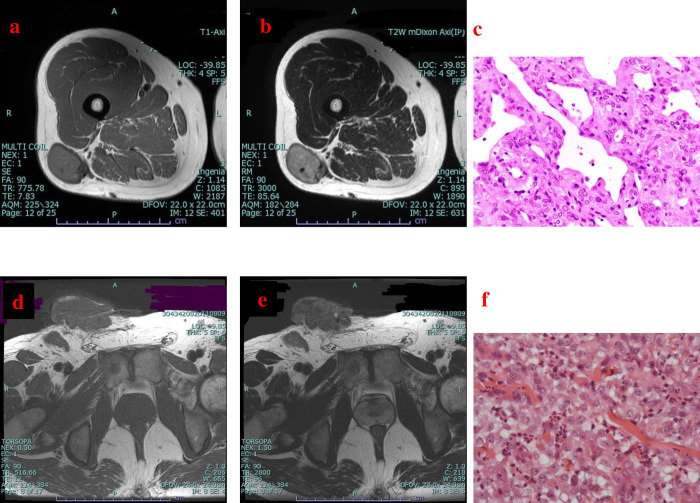
Fig 2. Representative cases.
(a, b, c) Magnetic resonance imaging (MRI) findings for a superficial lesion in the right thigh in a 44-year-old woman. Axial T1-weighted (a) and T2-weighted (b) images reveal that the lesion did not invade the skin. Pathological examination of the specimen confirmed a solitary fibrous tumor (c) (hematoxylin-eosin staining; magnification ×400). (d, e, f) MRI findings for a superficial lesion in the right inguinal region in a 46-year-old man. Axial T1-weighted (d) and T2-weighted (e) images reveal that the lesion invaded the skin. Pathological examination of the specimen confirmed epithelioid sarcoma (f) (hematoxylin-eosin staining; magnification ×400).

