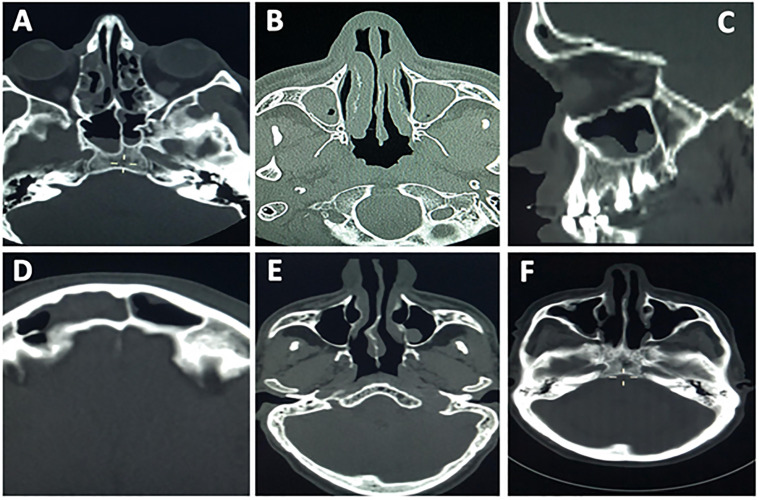
Fig 3.
Patients brain CT scans show the incidental detection of paranasal sinuses abnormalities; (A) right maxillary antrum polyp, (B) bilateral maxillary polyp with retention cyst, (C) maxillary antrum polyp and right nasal air fluid level likely abscess, (D) right frontal sinusitis, (E) normal paranasal study, (F) right maxillary antrum mucosal thickening.

