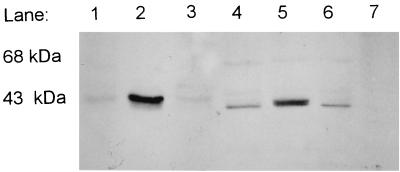
FIG. 1.
Western blot with anti-E1α serum (10) of crc mutants grown in 2xYT plus valine-isoleucine. The extracts in lanes 1 to 6 were from cultures grown in 2xYT plus 0.3% valine and 0.1% isoleucine (wt/vol); the extract in lane 7 was from a culture grown in glucose minimal medium. Each lane contained 5 μg of protein. Lane 1, P. putida PpG2; lane 2, P. putida JS394; lane 3, P. putida JS394(pJRS196); lane 4, P. aeruginosa PAO1; lane 5, P. aeruginosa PAO8020; lane 6, P. aeruginosa PA8020(pJRS196); lane 7, P. putida PpG2 grown in glucose. All cultures were grown to an A660 of between 0.6 and 0.8, and cell extracts were prepared as described before (16). Electrophoresis was done in an SDS–8.5% PAGE gel. Western blots were screened by using the ECL-Western blotting analysis system (Amersham Pharmacia Biotech) with Hybond-ECL nitrocellulose membranes according to the manufacturer's instructions. To determine whether the ECL detection method was quantitative, increasing amounts of PpG2 grown in valine-isoleucine-lactate medium were loaded on a gel and used for Western blotting with anti-E1α serum. The blot was scanned with a Molecular Dynamics densitometer, and pixel values versus micrograms of protein were graphed and shown to be a linear plot (data not shown).

