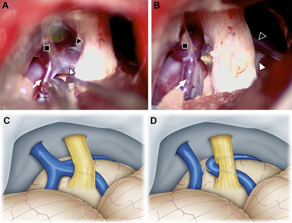
FIGURE 2.

Intraoperative photographs of a 67-year-old man who underwent microvascular decompression for right trigeminal neuralgia. A and C, The vein of the cerebellopontine fissure (white arrowhead) penetrates the trigeminal nerve, the transverse pontine vein (black arrowhead) contacts the ventral side of the trigeminal nerve, and the trigeminal nerve is in traction. B and D, By cutting a part of the superior petrosal vein (SPV, black square), the blood flow from the pontotrigeminal vein (white arrow) to the SPV was maintained, and the blood flow from the vein of cerebellopontine fissure to the SPV was diverted to the transverse pontine vein. SPV, superior petrosal vein.
