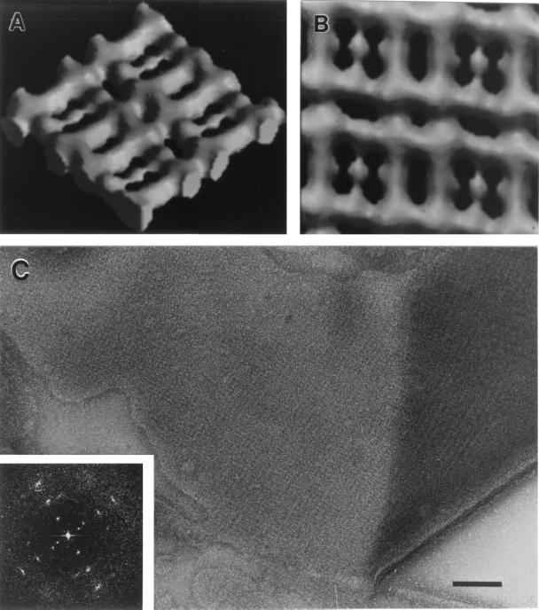
FIG. 3.
Structure of the oblique S-layer of P. uncinatum (54): Computer-generated side (A) and top (B) view of a surface-shaded, solid three-dimensional model of one side of the S-layer. According to the distribution of the measured data in Fourier space, the resolution of the reconstruction used for the model was about 2.0 nm. The lattice constants of the S-layer are a is 10 nm, b is 9.6 nm, and γ is 97.5°. (C) Appearance of the P. uncinatum cell wall after mechanical preparation of the S-layer (also see reference 56). The inset shows the corresponding optical diffraction pattern (power spectrum) which has been used for the reconstruction of the S-layer.

