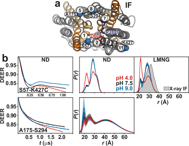
Fig. 4. Enhanced periplasmic flexibility of HnSpns by protonation, with a lateral opening between TMs 5 and 8.

a Spin label pairs (blue spheres) on the gating helices across the NTD and CTD are depicted on the periplasmic side. b Raw DEER decays and fits (left) are presented for the distance distributions P(r) in lipid nanodiscs (middle) and in LMNG micelles (right). The distributions predicted from the IF crystal structure are shaded gray.
