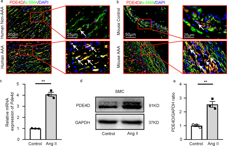
Fig. 2. PDE4D is primarily expressed in smooth muscle cells of AAA tissues.
a Immunofluorescence staining of PDE4D and α smooth muscle actin (α-SMA, smooth muscle cell marker) in human non-AAA sections and AAA tissues (6 μm). We obtained similar results from at least three different sets of human tissues in separate experiments. PDE4D: red, α-SMA: green. b Immunofluorescence staining of PDE4D and α-SMA in mouse control sections and AAA tissues (6 μm). We obtained similar results from at least three different sets of mouse samples in separate experiments. PDE4D: red, α-SMA: green. L: lumen. c RT-PCR analysis of Pde4d expression in rat aortic SMCs treated with Ang II (100 nM, 24 h). **p < 0.0001, Welch’s t test, mean ± SEM, n = 3 separate experiments. d Representative immunoblot analysis of PDE4D protein expression in SMCs treated with Ang II (100 nM, 24 h). e Quantification of PDE4D protein expression by immunoblotting in (d) normalized to GAPDH protein (fold change versus control). **p < 0.01, unpaired Student’s t test, mean ± SEM, n = 3 separate experiments.

