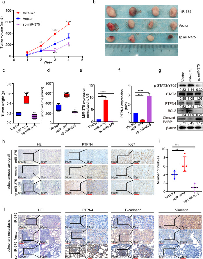
Fig. 5. MiR-375 overexpression promoted tumor growth and metastasis in vivo.
a Tumor volume during follow-up for 4 weeks. **p < 0.01, ***p < 0.001, ****p < 0.0001 vs. vector group. b Representative images of tumors in nude mice at the end timepoint. c Final tumor weight, d final tumor volume, e the expression of miR-375, and f mRNA level of PTPN4 were determined by qRT-PCR in tumor tissues at the endpoint of follow-up. g Western blotting was performed to determine the expression of p-STAT3, PTPN4, BCL2 and cleaved-PARP, and h IHC analysis of the expression of PTPN4 and Ki67 in DU145 tumor tissues as the expression of miR-375 was manipulated. i Quantification of the metastatic nodules calculated from the discontinuous lung sections. j HE staining and IHC analysis of the expression of PTPN4, E-cadherin and Vimentin using metastatic lung tissue. Vector: DU145 cells stably transfected with pSUPER-RETRO-Puro-empty vector and pHB-U6-MCS-PGK-PURO-empty vector. MiR-375: DU145 cells stably transfected with pSUPER-RETRO-Puro-miR-375 vector and pHB-U6-MCS-PGK-PURO-empty vector. Sp miR-375: DU145 cells stably transfected with pHB-U6-MCS-PGK-PURO-miR-375 sponge vector and pSUPER-RETRO-Puro-empty vector.

