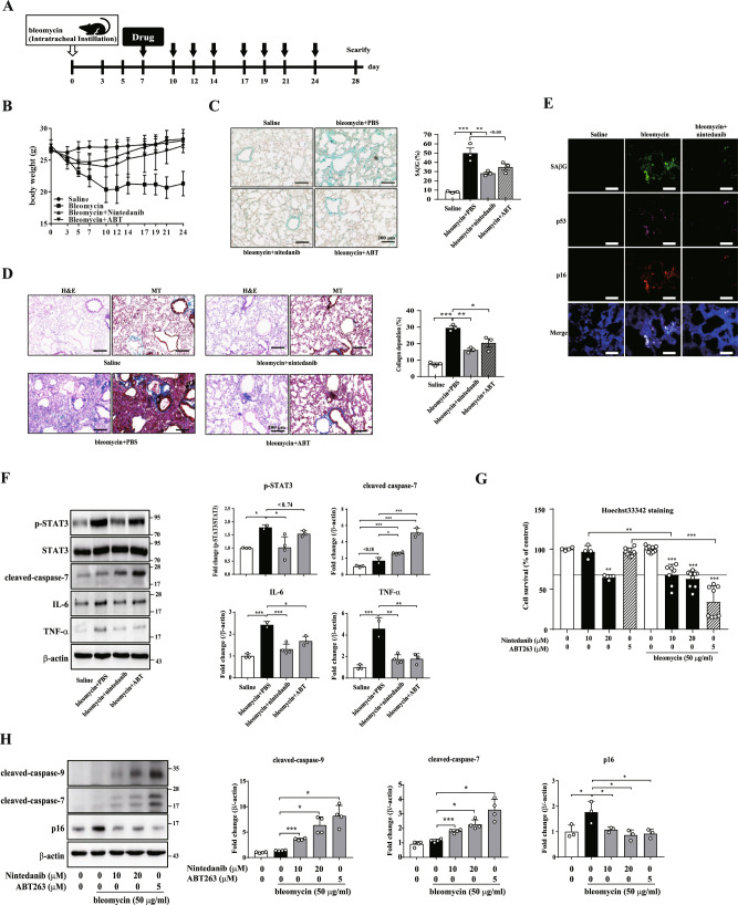
Fig. 5. Nintedanib shows senolysis in bleomycin-induced in vivo and in vitro model.
A Experimental scheme for establishing bleomycin-induced lung fibrosis model: saline (n = 3), PBS (bleomycin; n = 2), ABT263 (bleomycin + ABT; n = 3), or nintedanib (bleomycin + nintedanib; n = 4) was administrated via intraperitoneal injection three times per week for 24 days (black arrow). Control mice were treated with PBS are labeled saline (n = 3). B Body weight changes after bleomycin followed by drug treatment. C, D Lung tissues were stained with an SAβG staining kit to measure the number of senescent cells (C), hematoxylin and eosin (H&E) to assess lung interstitial damage, and Masson’s trichrome (MT) staining (collagen is stained blue) to detect collagen deposition (D). Scale bar, 200 μm. E Lung tissues were stained with SPiDER-βGal to measure SAβG activity and with anti-p16 and anti-p53 antibodies to determine whether senescence was attenuated. Scale bar, 200 μm. F Western blot assays using anti-p-STAT3, anti-STAT3, anti-caspase-7, anti-IL-6 and anti-TNF-α antibodies were performed to identify the apoptotic pathways involved in the lung tissue. G Bleomycin-induced senescent HPFs was treated with nintedanib or ABT263 for 3 days. Then, Hoechst 33342 staining was performed to assess cell viability. n = 3 H Bleomycin-induced senescent HPFs were treated with nintedanib or ABT263 for 3 days, and then, western blot assays using anti-caspase-9, anti-caspase-7, and anti-p16 antibodies were performed to identify the apoptotic pathways involved in the senolytic effect. The data normalized to those for DMSO-treated cells are shown as the mean ± S.D (F, G). *p < 0.05, **p < 0.01, ***p < 0.001 by one-way ANOVA with Tukey’s post hoc test.

