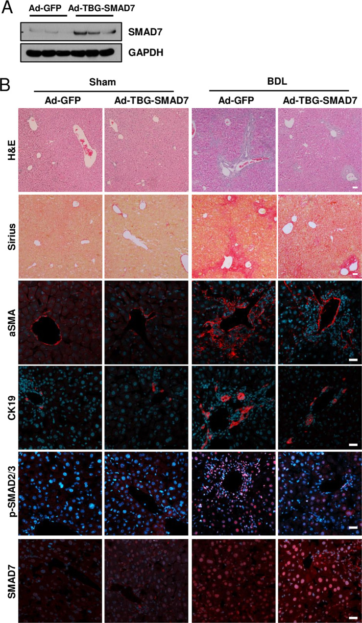
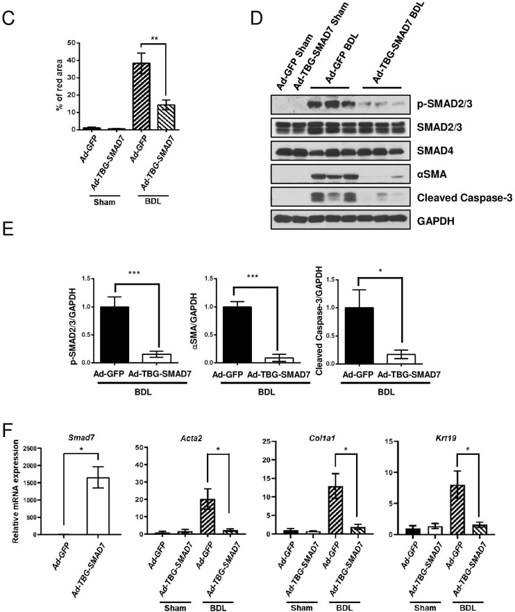
Fig. 7. Hepatocyte-specific overexpression of SMAD7 in liver-specific Prom1-deficient mice ameliorated BDL-induced liver fibrosis.
Eight-week-old f/f; Alb-Cre mice were infected by adenovirus expressing GFP or TBG-SMAD7. The mice underwent sham operation (n = 3) or BDL (n = 5–9). A SMAD7 expression in the liver was determined by immunoblotting 3 days after BDL. B Seven days after BDL, the liver was analyzed by H&E and Sirius Red staining and immunofluorescence analysis of αSMA and CK19, p-SMAD2/3, and SMAD7. C Liver fibrosis was quantified by measuring the area stained with Sirius Red. Two to three images of each liver showing Sirius Red staining were obtained. D Each specimen was analyzed by immunoblot analysis of p-SMAD2/3, SMAD2/3, SMAD4, αSMA, cleaved Caspase-3, and GAPDH. E The band intensities of p-SMAD2/3, αSMA, and cleaved Caspase-3 in D were statistically analyzed after normalization to the band intensity of GAPDH. F The mRNA levels of Smad7, Acta2, Col1a1, and Krt19 in each liver specimen were analyzed by RT–qPCR. The mRNA levels were normalized to those of 18S rRNA. TBG, thyroxine-binding globulin. Scale bar = 20 µm. *p < 0.05, **p < 0.01, ***p < 0.001. All data are the mean ± S.E.M.


