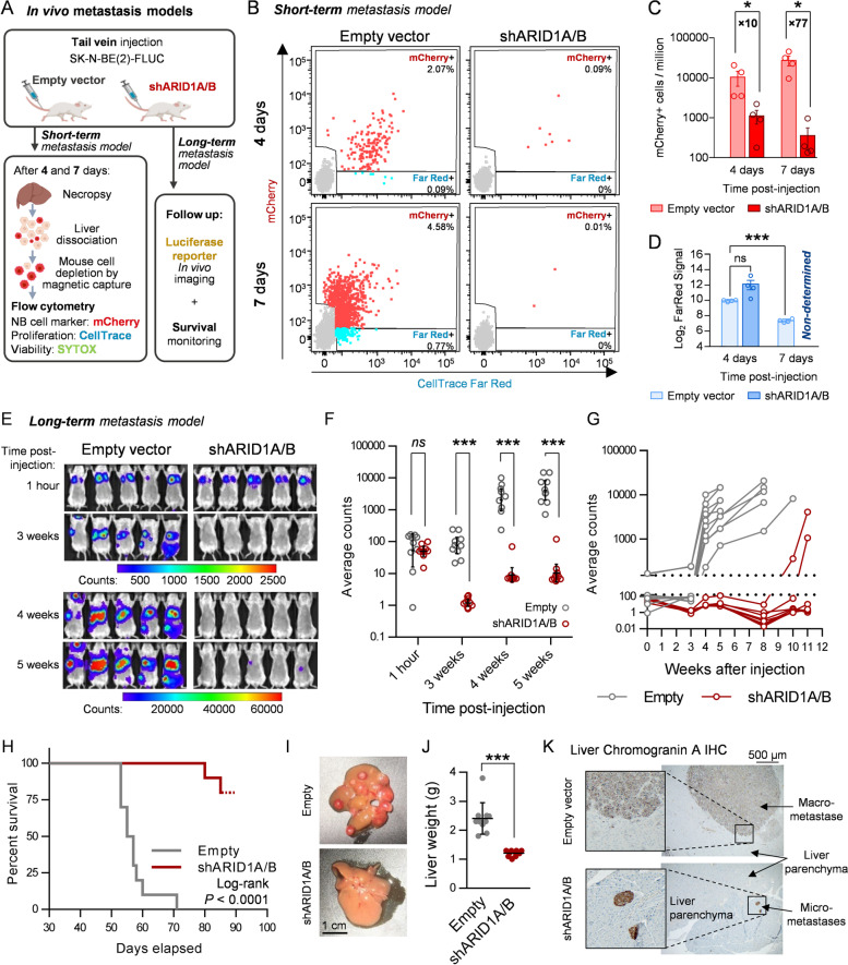
Fig. 5.
BAF disruption impairs neuroblastoma metastasis initiation and growth in vivo. A Experimental design of the in vivo short- and long-term neuroblastoma metastasis models performed with SK-N-BE(2) cells. B Representative flow cytometry plots of the short-term metastasis experiment, showing mCherry positive (mCherry +) and FarRed positive/mCherry negative (FarRed +) single living cell populations. C Quantification of detected mCherry positive cells, expressed in events per million of living single cells (parent gate). Mann–Whitney’s test was performed for statistical comparisons. Fold change between conditions are indicated. D Average FarRed intensities assessed, when possible, for the mCherry + population of each experimental group. E Representative in vivo luminescence images of 5 mice per group at the indicated time points. Scale bar represents luminescence counts (photons). F Luminescence quantification, expressed in average counts, and comparison between experimental groups at the indicated times post-injection. G Individual mice luminescence quantification follow-up through the entire experiment. H Kaplan–Meier survival plot comparing mice injected with SK-N-BE(2) transduced with empty vector (Empty) or shARID1A/B. Log-rank test was performed to assess statistical significance. I Representative images of mouse livers after necropsy. (J) Comparison between groups of liver weight at necropsy. K Representative bright field microscopy images of chromogranin A immunohistochemistry of FFPE liver slides. ns means ‘non-significant’; * means p < 0.05; ** means p < 0.01; *** means p < 0.001

