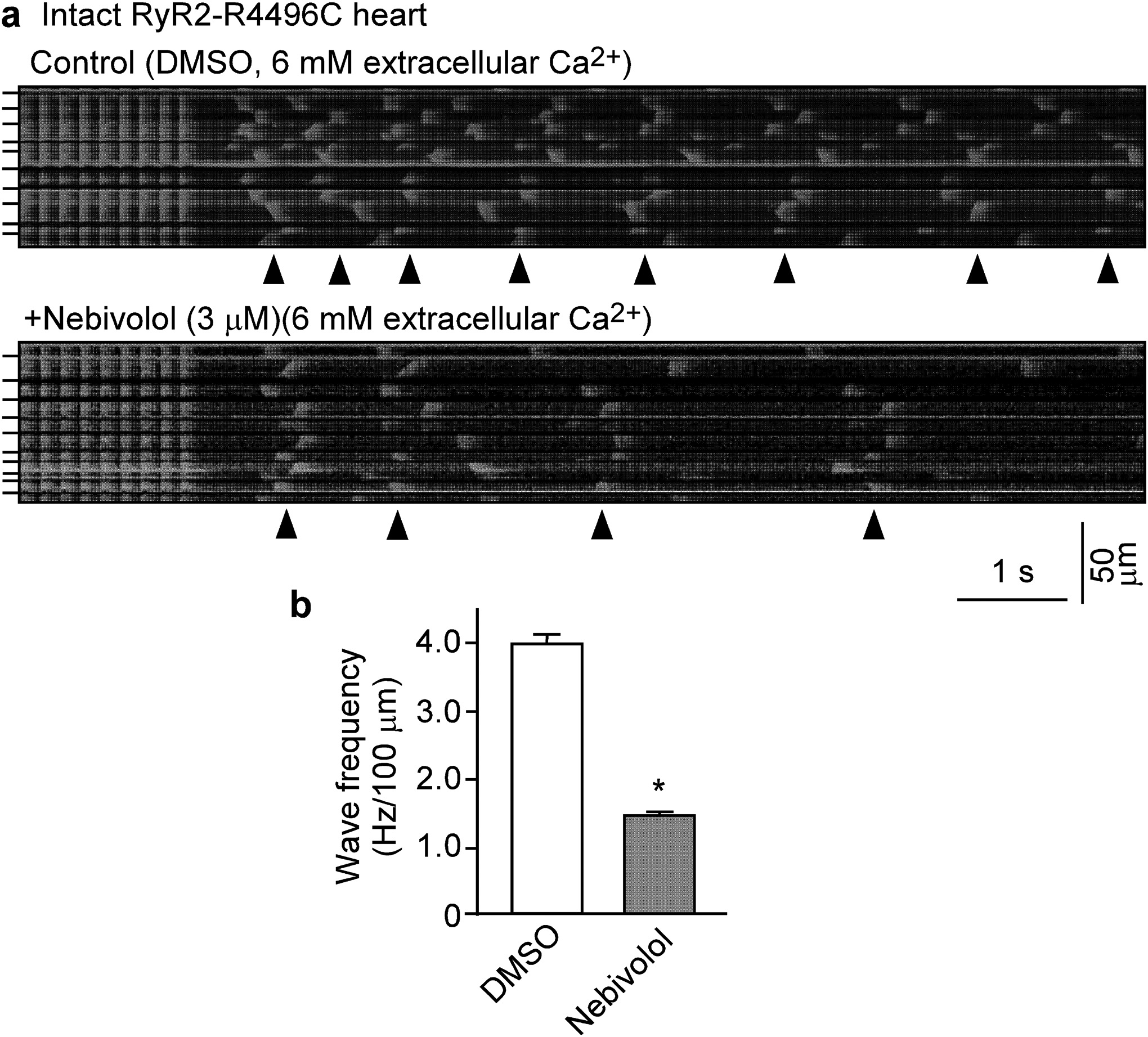
Figure 4. Nebivolol suppresses Ca2+ waves in intact hearts.

Intact hearts from heterozygous RyR2-R4496C mutant mice were isolated and loaded with Rhod-2 AM, and Langendorff-perfused with KH solution containing 6 mM extracellular Ca2+ to induce SR Ca2+ overload. AV node was ablated by electro-cautery and the hearts were paced at 6Hz, then pacing was stopped. (a) Representative line-scan confocal images of Ca2+ dynamics in hearts 2 hours after perfused with DMSO (control, top panel) or with nebivolol (3μM, bottom panel). (b) Frequency of spontaneous Ca2+ waves. Arrowheads show the occurrence of Ca2+ waves. Short bars to the left indicate cell boundaries within the intact heart. Data shown are means ± SEM from 16–18 scan areas of 3 hearts for each group. (*P < 0.01 vs control).
