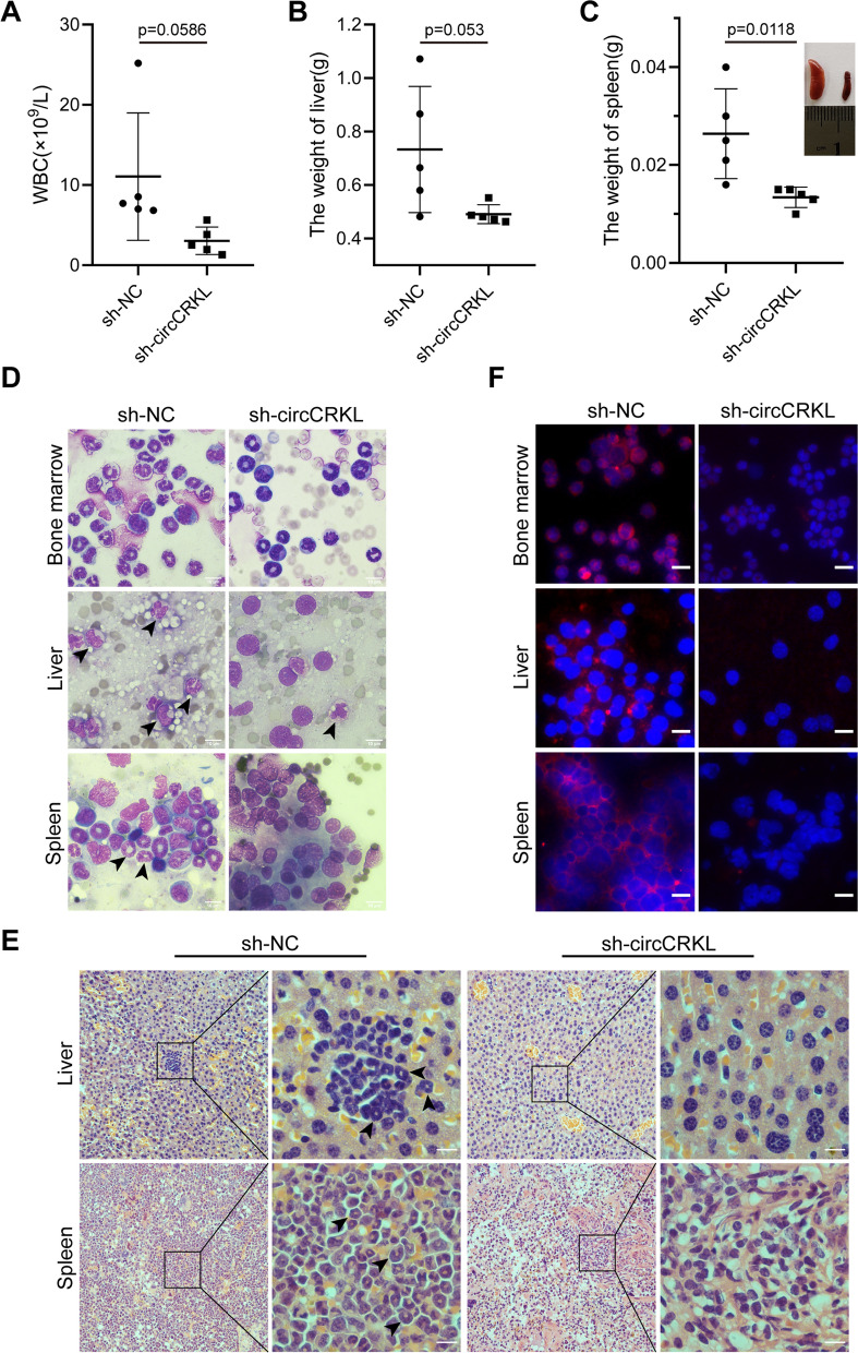
Fig. 3.
circCRKL enhances CML cells proliferation in vivo. A The number of WBCs was calculated. B, C The weights of liver and spleen were recorded and the images were shown. D Wright’s staining was performed to observe the leukemic cells in bone marrow, liver, and spleen, the leukemic cells were indicated by the black arrow. Scale bar, 10 μm. E The leukemic infiltration in liver and spleen was observed by H&E staining. F BCR-ABL levels in bone marrow, liver, and spleen cells were determined with immunofluorescence. Scale bar, 10 μm. *p < 0.05 and ** < 0.01

