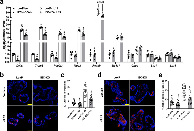
Fig. 3. Loss of SIRT6 in intestinal organoids causes defective IL13-induced tuft and goblet cell hyperplasia.
LoxP and IEC-KO intestinal organoids were treated with vehicle or rIL13 (25 ng/ml) for 48 h and then subjected to the following analyses. a qPCR analysis of IEC markers expression. n = 4 biological replicates/group; from left to right, Dclk1 (*p = 0.0001, 0.0014, 0.0001; #p = 0.0012, 0.01); Trpm5 (*p < 0.0001, <0.0001, 0.0018; #p < 0.0001, 0.0015); Pou2f3 (*p = 0.0303, 0.0018, <0.0001; #p = 0.0018, 0.017); Muc2 (*p < 0.0001, 0.002; #p < 0.0001, 0.0154); Retnlb (*p = 0.0087, 0.0013; #p = 0.0087); Slc5a1 (*p = 0.0019, 0.0116; #p = 0.0032); Lyz1 (*p = 0.049, 0.0339; #p = 0.0008); Lgr5 (*p = 0.0265, 0.0326; #p = 0.0082). b, d Tuft and goblet cells were labeled by anti-DCLK1 (b) and anti-MUC2 (d), respectively, in frozen sections of organoids (200X). c Quantification of tuft cells shown in (b). n = 15 organoids/group; from left to right, *p = 0.0301, <0.0001, 0.001; #p < 0.0001 for both. e Quantification of goblet cells shown in (d). n = 15 organoids/group; from left to right, *p < 0.0001, 0.0176; #p < 0.0001, 0.0032. Data are presented as mean ± SEM. All p values were generated by two-tailed unpaired t test. *p < 0.05 vs LoxP-Veh, #p < 0.05 vs LoxP-rIL13. Scale bars, 50 µm. Source data are provided as a Source Data file.

