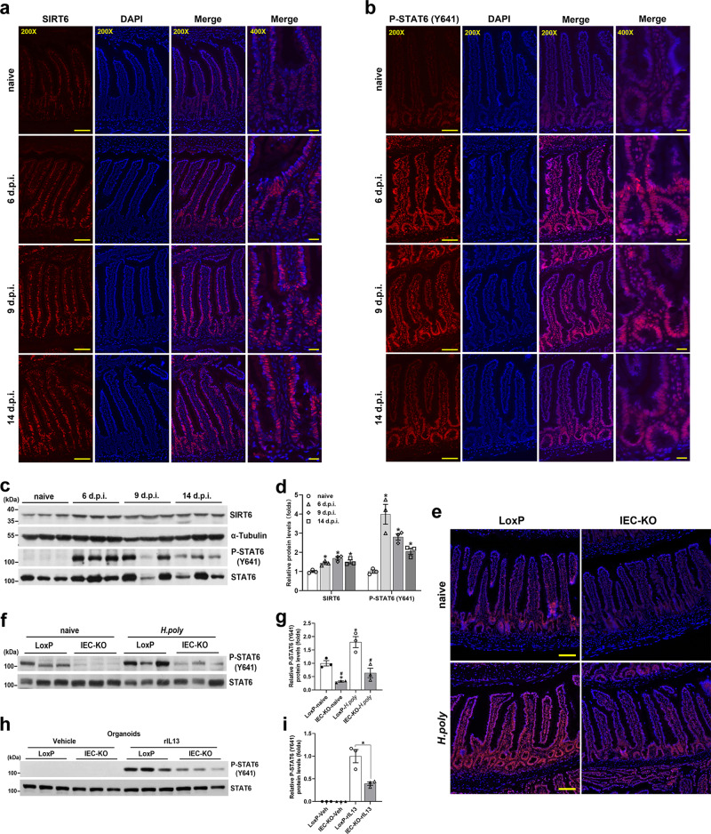
Fig. 4. IEC ablation of Sirt6 attenuates the Tyr641 phosphorylation of STAT6.
a–d 2-month-old C57BL/6J male mice were infected with H. poly and analyzed on indicated days post-infection. a, b Immunostaining for SIRT6 (a) and P-STAT6 (Y641) (b) in the jejunum. c, d Western blot (c) and quantification (d) analyses of SIRT6 and P-STAT6 (Y641) in the jejunal IECs. n = 3 mice/group; p = 0.0102 (6 dpi), 0.006 (9 dpi), 0.0203 (14 dpi) for SIRT6; p = 0.0259 (6 dpi), 0.0025 (9 dpi), 0.0089 (14 dpi) for P-STAT6. e–g 2–3-month-old male naive and H. poly-infected LoxP and IEC-KO mice were subjected to the following assays on day 14 post-infection. e Immunostaining for P-STAT6 (Y641) in the jejunum. f, g Western blot (f) and quantification (g) analyses of P-STAT6 (Y641) in the jejunal IECs. n = 3 mice/group; from left to right, *p = 0.0166, 0.0423; #p = 0.0177, 0.0136. h, i Western blot (h) and quantification (i) analyses of P-STAT6 (Y641) in vehicle- or rIL13-treated (25 ng/ml, 48 h) organoids. n = 3 biological replicates/group; p = 0.0451. Data are presented as mean ± SEM. All p values were generated by two-tailed unpaired t test. In d, *p < 0.05 vs naive; In g, *p < 0.05 vs LoxP-naive, #p < 0.05 vs LoxP-H. poly; Scale bars, 200X, 100 µm; 400X, 25 µm. Source data are provided as a Source Data file.

