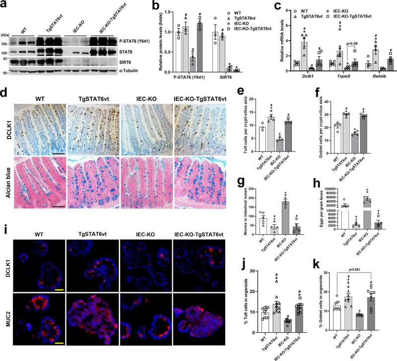
Fig. 6. IEC STAT6vt overexpression rescues the defective intestinal type 2 immunity resulting from Sirt6 deletion.
2–3-month-old male mice with 4 genotypes (WT, TgSTAT6vt, IEC-KO, IEC-KO-TgSTAT6vt) were infected with H. poly and subjected to the following analyses on day 14 post-infection. a, b Western blot (a) and quantification (b) analyses of P-STAT6 (Y641) and SIRT6 in the jejunal IECs. n = 3 mice/group; from left to right, P-STAT6 (*p = 0.0083; #p = 0.0074, 0.0026); SIRT6 (*p = 0.0358, 0.0344; #p = 0.0002). c qPCR analysis of the expression of Dclk1, Trpm5 and Retnlb in the jejunal IECs. Dclk1, Trpm5, n = 5 mice/group; Retnlb, n = 4 mice/group; from left to right, Dclk1 (*p = 0.0054, 0.0019; #p = 0.0027, 0.0494); Trpm5 (*p = 0.0389, 0.0085; #p = 0.0184); Retnlb (*p = 0.0466; #p = 0.0232, 0.0154). d Tuft and goblet cells were examined by DCLK1 immunostaining and Alcian blue staining, respectively in the jejunum (200X). e, f Quantification of tuft (e) and goblet (f) cells shown in (d). n = 3, 4, 3, 3 mice/group, respectively in (e) and n = 4 mice/group in (f); 30 crypt-villus units counted for each mouse; form left to right, *p = 0.0253, 0.0119; #p = 0.0007, 0.0017 in (e); *p = 0.0059, 0.0264, 0.0104; #p < 0.0001 for both in (f). g, h Analysis of parasite burden by quantification of adult worms in intestinal lumen (g) and eggs in feces (h). n = 5, 4, 5, 4 mice/group; form left to right, *p = 0.0458, 0.0016; #p = 0.0001, 0.0005 in (g); *p = 0.0487, 0.0268; #p = 0.0051, 0.0052 in (h). i Tuft and goblet cells were labeled by anti-DCLK1 and anti-MUC2, respectively, in frozen sections of rIL13-treated organoids (200X). j, k Quantification of tuft (j) and goblet (k) cells shown in (i). n = 10 (j) and 9 (k) organoids/group; form left to right, *p = 0.0307, 0.0003, 0.0281; #p = 0.0003, <0.0001 in (j); *p = 0.0181, 0.0002; #p = 0.0002, 0.0009 in (k). Data are presented as mean ± SEM. All p values were generated by two-tailed unpaired t test. *p < 0.05 vs WT, #p < 0.05 vs IEC-KO. Scale bars, 100 µm in (d); 50 µm in (i). Source data are provided as a Source Data file.

