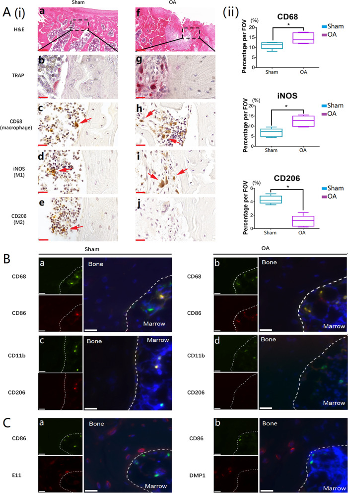
Fig. 4.
Macrophages were actively involved in inflammatory bone remodeling and M1 macrophages were the major activated macrophage subtype in OA bone remodeling areas. A(i) H&E, TRAP, and IHC staining of macrophage specific markers (CD68: macrophage pan marker; iNOS: M1 macrophage marker; CD206: M2 macrophage marker; positive cells were labeled with red arrows) on normal and OA bone sections (a and f: scale bars represented was 200 μm in H&E staining; b and g: scale bars represented was 20 μm in TRAP staining; c, d, e, h, i, and j: scale bars represented was 20 μm in IHC staining); A(ii): The box plot graphs demonstrated the positive cells per field of view. Data was shown as the mean ± SD (*p < 0.05, t-test); B IF double staining of macrophage markers on normal and OA bone sections (CD68 and CD11b: macrophage pan markers; CD86: M1 macrophage marker, CD206: M2 macrophage marker; scale bars represented 20 μm); C IF double staining of M1 macrophage marker and osteocyte marker on OA bone sections (CD86: M1 macrophage marker; E11: early osteocyte marker; DMP1: mature osteocyte marker; scale bars represented 20 μm)

