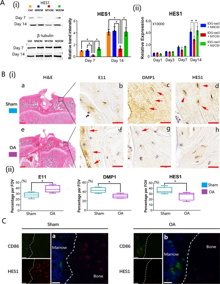
Fig. 5.
Notch signaling pathway was inhibited in M1 macrophage-stimulated osteocytes and Notch signaling pathway was downregulated in OA inflammatory bone remodeling areas. A(i) Expression of HES1 from macrophage-derived medium cultured osteocytes detected by western blot; A(ii) qRT-PCR measurement of HES1 gene expression from macrophage-derived medium cultured osteocytes. Data from three independent experiments performed under the same condition were shown as mean ± SD (*p < 0.05, one-way ANOVA); B normal and OA bone sections stained with H&E and IHC (E11: immature osteocyte marker; DMP1: mature osteocyte marker; HES1: Notch signaling pathway marker; a and e: scale bar present was 100 μm; b, c, d, f, g, and h: scale bar present was 20 μm); C IF double staining images of normal and OA bone sections (CD86: M1 macrophage marker; HES1: Notch signaling pathway marker; the scale bars present was 20 μm)

