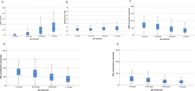
Fig. 2.
Distribution of small airway abnormality indicated by markers from CT, IOS and spirometry with age stratification in total subjects. Panel A was for LAA·856 from CT. Panel B was for R5–R20 from IOS. Panel C was for MMEF, %predicted from spirometry. Panel D was for FEF·50, %predicted from spirometry. Panel E was for FEF·75, %predicted from spirometry. Note: Abbreviations: LAA-856 = low-attenuation area of the lung with attenuation values below -856 Hounsfield units on full-expiration CT; R5–R20 = the difference from resistance at 5 Hz to resistance at 20 Hz; MMEF, %predicted = maximal mid-expiratory flow of percent predicted; FEF50, %predicted and FEF75, %predicted = forced expiratory flow at 50 and 75 of forced vital capacity of percent predicted

