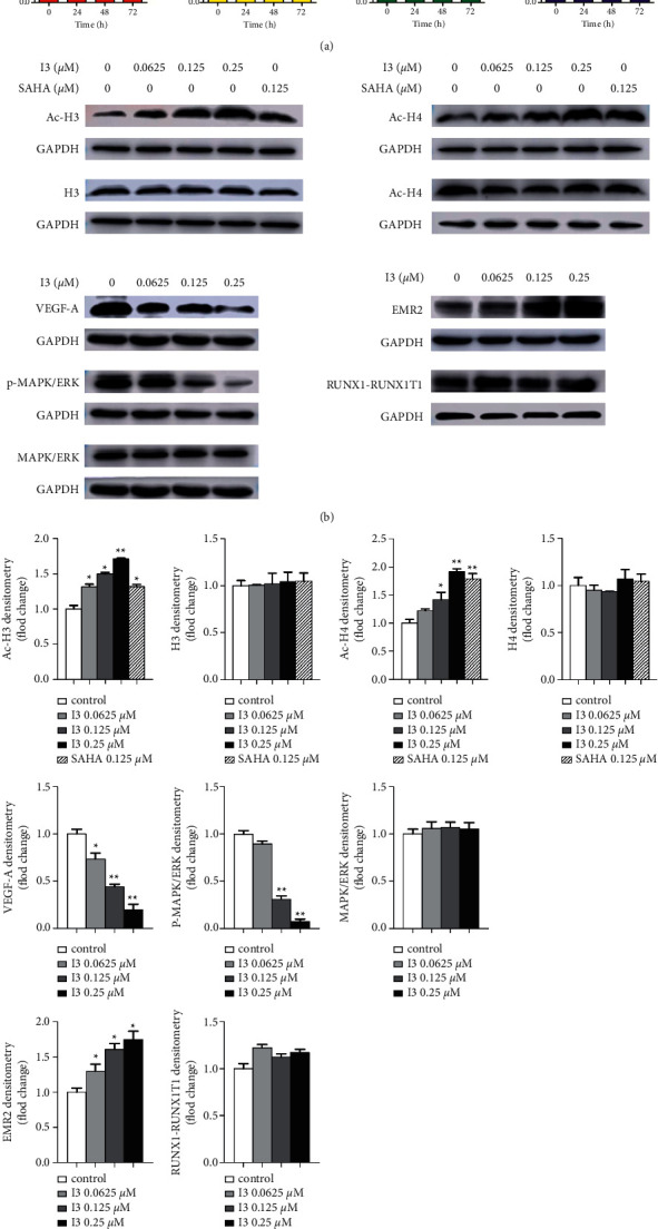
Figure 7.

The RT-PCR and western blotting analysis of cell differentiation-related genes and proteins in Kasumi-1 cells. (a) The effect of I3 on the mRNA expression of RUNX1-RUNX1T1, ERK, VEGF-A, and EMR2 measured by RT-PCR. (b) The effect of I3 on the protein expression of H3, Ac-H3, H4, Ac-H4, VEGF-A, MAPK/ERK, p- MAPK/ERK, RUNX1-RUNX1T1, and EMR2 measured by western blotting analysis. (c) Graph bars show the protein expression quantified by the AI600 imager. Cells were incubated with 0.25 μM of I3 for 72 h (∗p < 0.05, ∗∗p < 0.01).
