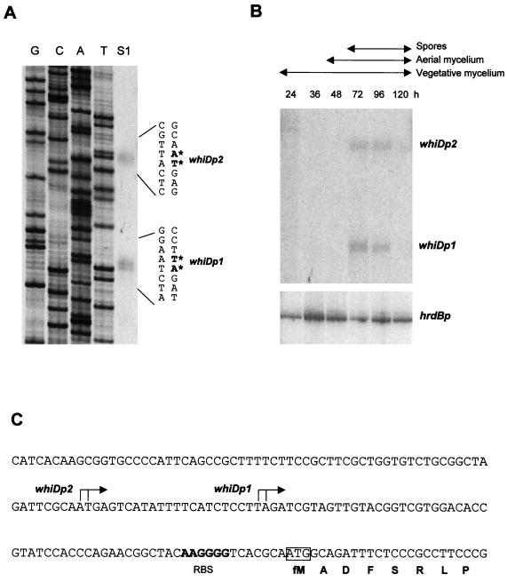
FIG. 7.
Transcriptional analysis of whiD. (A) High-resolution S1 nuclease mapping of the 5′ ends of the whiDp1 and whiDp2 transcripts. The lane labeled “S1” represents the DNA fragments protected by RNA initiating from whiDp1 and whiDp2. The most likely transcription start points are indicated by the asterisks. Lanes labeled G, C, A, and T represent a dideoxy sequencing ladder generated by using the same oligonucleotide that was used to generate the S1 mapping probe. The RNA used was from the 72-h time point from panel B. (B) S1 nuclease protection analysis of transcription from whiDp1, whiDp2, and hrdBp during development. RNA was isolated from wild-type S. coelicolor grown on cellophane discs on MM containing mannitol as carbon source. The time points (in hours) at which mycelium was harvested for RNA isolation, and the presence of vegetative mycelium, aerial mycelium, and spores as judged by microscopic examination, are shown. The hrdB panel from this figure was published previously (25, 26, 39) and is shown here for comparison with the whiD data. (C) Nucleotide sequence of the whiD promoter region indicating the whiDp1 and whiDp2 transcription start points, the putative ribosome binding site (RBS), and the start of the whiD coding sequence.

