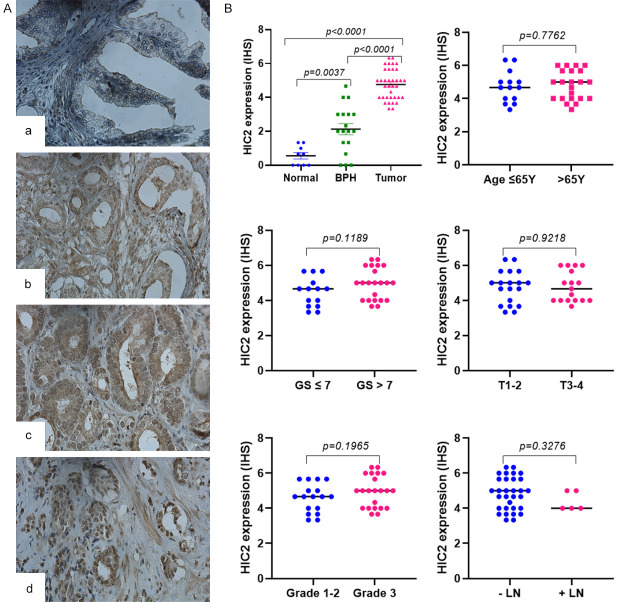
Figure 6.
Protein expression of HIC2 in PCa tissue specimens. HIC2 expression was evaluated by immunohistochemistry in tissue microarray slide comprising 50 tissue cores of PCa, 20 BPH, and ten normal prostate tissues. A: Immunohistochemical staining of HIC2 in tissues collected from normal individuals (a) and PCa tissue cores at different Gleason scores (GSs): b (GS<7), c (GS=7) and d (GS>7). B: Quantification of HIC2 expression in PCa tissues considering age, Gleason score, pathological stage, tumor grades, and lymph node involvement. Significance of the data was calculated at P<0.05. Magnification is 400X.

