
Figure 1.
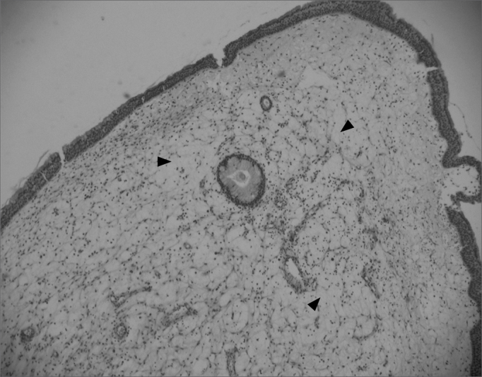
Edematous or eosinophilic polyp in a panoramic view. We can see the edema in the region of the stroma, with broad intercellular spaces (arrows). The stromal edema is partially filled by intercellular fluid, forming pseudocystic spaces (H&E × 40).