
Figure 4.
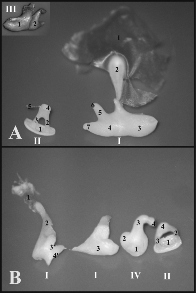
A) Set of ossicles of a young guinea pig. Detailed view of the malleus-incus joint (III) (microphotography, 58X magnification); B) Set of ossicles from a young-adult rat (microphotography, 60X magnification). I - Malleus - Incus 1 - Tympanic membrane; 2 - Malleus handle; 3 - Head of the malleus; 3’ - Neck of the malleus; 4 - Incus (fused to the malleus); 4’ - Joint surface of the malleus-incus; 5 - Long process of the incus; 6 - Lenticular process of the incus; 7 - short process of the incus II - Stapes 1 - Footplate of the incus; 2 - anterior crus of the stapes; 3 - Stapes posterior crus; 4 - Stapes Capitulum; 5 - Stapes muscle tendon III - Malleus-incus joint 1 - Malleus; 2 - Incus IV - Incus 1 - Incus-Malleus joint surface; 2 - Short process of the incus; 3 - Long process of the incus; 4 - Lenticular process of the incus.