
Figure 8.
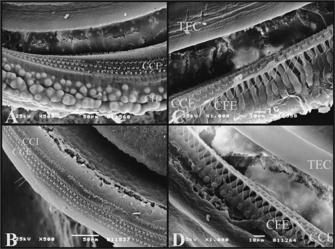
A) 3rd cochlear turn of the guinea pig showing the Hansen Cells (CH) (scanning electron micrograph, 500X magnification); B) 2nd cochlear turn from a rat, showing the lack of Hansen Cells definition (Scanning electron microscopy, 500X magnification); C) Guinea pig’s cochlea basal turn showing the external phalanx cells (CFE) (Deiters) (scanning electron micrography, 1000X magnification); D) Rat’s cochlea basal turn showing the external phalanx cells (CFE) (Deiters) (Scanning electron micrography, 1000X magnification).