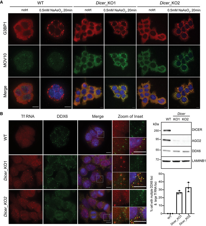
Figure 2. Cytosolic L1 RNP foci are poised to be stress granules that co‐localize with multiple small DDX6 foci.

-
AWT and Dicer_KO mESCs were treated with 0.5 mM sodium arsenite (NaAsO2) for 20 min or left untreated prior to fixation with formaldehyde. Maximum intensity projections across Z stacks of example images from indicated mESCs immunostained for G3BP1 (red) and MOV10 (green) with nuclei stained with DAPI (blue).
-
BRepresentative Western blots showing low AGO2 protein levels in Dicer_KO as compared to WT mESCs (right side). LAMINB1 served as loading control. On the left, maximum intensity projections across Z stacks of example images from indicated mESCs stained for L1 Tf RNA FISH (red) combined with immunostaining for a resident protein of P‐bodies, DDX6 (green) and nuclei stained with DAPI (blue). The gray square marks position of the inset. Yellow arrow heads point to cytoplasmic foci where L1 RNA and DDX6 protein co‐localize.
Data information: (A and B) are representative images of three independent experiments. Bar graphs represent mean ± SD. 94–150 cells per cell line were analyzed for each experiment. Scale bar 5 μm.
Source data are available online for this figure.
