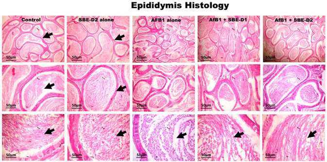
Figure 8.
Histology section from the epididymis of the control and SBE alone treated rats show typical morphology. The tubules are essentially normal with abundant spermatozoa (black arrows). AFB1 (50 µg/kg) alone treated rats’ epididymis presented reduced epididymal sperm cells. Groups treated with AFB1 and graded doses of—5 and 10 mg/kg body weight—of SBE show normal epididymis morphology. The tubules are essentially normal with SBE dose-dependent spermatozoa increases in the lumen (black arrows). (H&E-stained tissue sections; 1.08 cm = 50 µm). (A color version of this figure is available in the online journal.)

