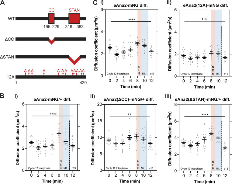
Figure 4.
Ana2’s change in diffusion rate does not appear to depend on the CC or STAN domain, but this change is perturbed if Ana2 cannot be phosphorylated by Cdk/Cyclins. (A) Schematic illustration of the Ana2 protein and the deletion/mutant forms analyzed in this study: central coiled-coil (CC) domain (aa195-229); STil/ANa2 (STAN) domain (aa316-383); the 12 S/T residues in S/T-P motifs that were mutated to Alanine. (B and C) Graphs show cytoplasmic FCS diffusion measurements (mean ± SEM) in embryos laid by females of the following genotypes: B (i) eAna2-mNG/+; B (ii) eAna2(∆CC)-mNG/+; B (iii) eAna2(∆STAN)-mNG; C (i) eAna2-mNG; C (ii) eAna2(12A)-mNG. Measurements were taken every 2 min from the start of nuclear cycle 12. The timing window of NEB is depicted in red and mitosis in blue. Each data point represents the average of 4–6× 10-s recordings from an individual embryo (N ≥ 13). Statistical significance was assessed using a paired one-way ANOVA test (for Gaussian-distributed data) or a Friedman test (****, P < 0.0001; **, P < 0.01).

