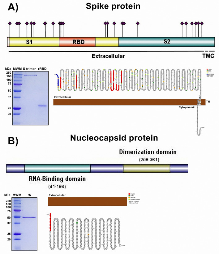
Figure 2.
Recombinant proteins of the spike glycoprotein and nucleocapsid of SARS-CoV-2 were obtained, and the primary and secondary structures were analyzed. (A) Spike protein. Upper panel and lower right panel, analysis of the primary and secondary amino acid sequences showing the extracellular, TM (transmembrane) and cytoplasmic (C) localization of each amino acid. Purple diamonds represent glycosylated residues. (B) N protein. Upper panel and lower right panel showing analysis of the primary and secondary amino acid sequences with intraviron localization, phosphorylated amino acids, RNA-binding and dimerization domains. In both proteins, selected peptides are shown in red in 2D structure, and the peptide signal is marked in blue. Middle panel in (A) and (B): electrophoresis and Coomassie blue staining of recombinant RBD, S trimer, or N. MWM molecular weight marker.

