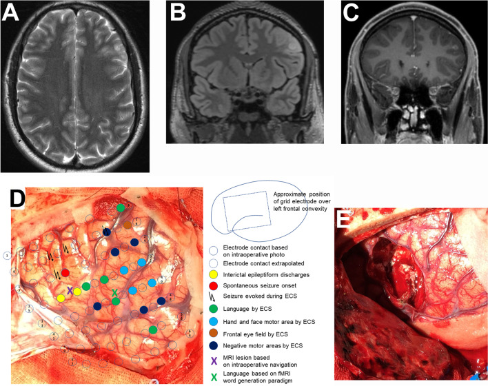
Fig. 2.
Brain tumor in the left middle frontal gyrus with cortical and subcortical T2/FLAIR hyperintense signal (A, B) and partial Gadolinium enhancement (C) in a 24-year-old male patient. A Axial T2, B coronal FLAIR, C coronal T1 with Gadolinium, D intraoperative situs with superimposed results from extraoperative subdural electrode recording and direct cortical stimulation, E intraoperative situs following resection of the tumor and adjacent epileptogenic area (histology: angiocentric glioma WHO grade 1).
Figure modifed from Wehner et al. [85]

