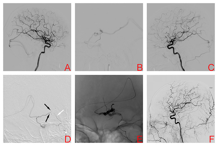
Figure 1.
Digital subtraction angiography left internal carotid artery (ICA) demonstrated the left basal ganglia arteriovenous malformation in case 1 (A). Superselective arteriography and embolization via the anterior choroidal artery (B). Immediate angiography of left ICA after transarterial embolization showed a residual small nidus (C). TVE was performed due to the lack of optimal arterial access. Dual microcatheters (black arrows) were positioned in the origin of the venous collector and a balloon microcatheter (white arrow) was placed in the ipsilateral internal carotid artery (D). After the balloon was inflated to provisionally occlude the internal carotid artery, we used the PCT to occlude the nidus (E). Digital subtraction angiography 4 months later confirmed AVM obliteration (F).

