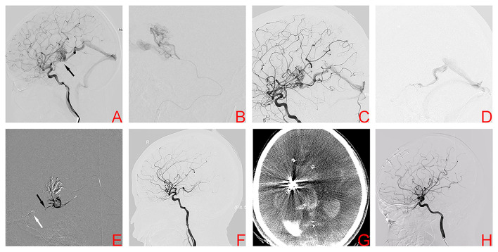
Figure 2.
Digital subtraction angiography demonstrates the right basal ganglia arteriovenous malformation in case 2 (A). Superselective arteriography and embolization via the posterior communicating artery (B). Angiography after transarterial embolization shows a partially embolized arteriovenous malformation (C). TVE was performed due to the lack of optimal arterial access. Transvenous microcatheter injection angiography confirmed an optimal position of the microcatheter tip (D). After the balloon (white arrow) was inflated to provisionally occlude the ipsilateral internal carotid artery, Onyx was injected transvenously into the AVM nidus through the microcatheter, and ~2 cm of embolysate reflux (black arrow) was encountered prior to achieving sufficient retrograde nidal penetration (E). Postoperative angiography showed complete embolization (F). However, postoperative CT confirmed intracranial hemorrhage (G). Angiography 1 year later showed complete embolization of the arteriovenous malformation (H).

