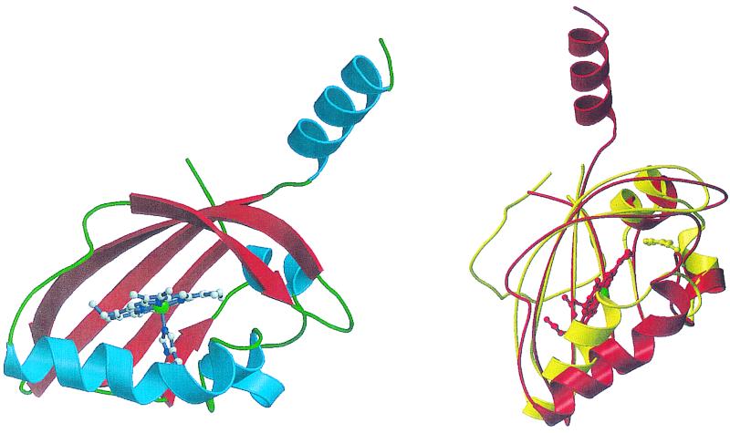
FIG. 2.
Structure of the heme-binding PAS domain from the FixL protein of Bradyrhizobium japonicum. (Left) A ribbons diagram with α-helices in blue and β-sheets in red. The helix pointing up leads to the HK domain. The heme moiety, with the heme iron in green, and its axial histidine ligand are shown. (Right) A comparison of the structures of the FixL heme-binding PAS domain in red and the PAS domain of the photoactive yellow protein PYP in yellow. The locations of the heme moiety of FixL, with the heme iron in green, and the p-hydroxycinnamate chromophore of PYP are indicated. Reprinted from reference 10 with permission of the publisher. (Figure provided by M.-A. Gilles-Gonzalez.)

