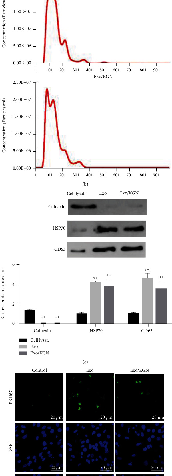
Figure 2.

Extraction and characterization of EXOs. (a) The morphology of EXOs in the Exo and Exo/KGN groups was observed by TEM. (b) The diameters of EXOs were analyzed with NTA software. (c) The protein expression levels of Calnexin, HSP70, and CD63 in cell lysates and Exos were detected by Western blot. (d) The level of EXO uptake by ADSCs was detected through PKH67 labeling. ADSC nuclei were stained with 4',6-diamidino-2-phenylindole (DAPI). ∗∗P < 0.01 vs. cell lysate group.
