
Figure 1.
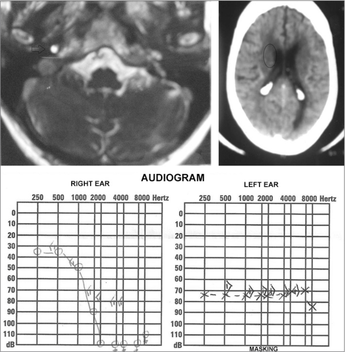
Complementary tests. Upper left-hand image: MRI showing an acoustic neuroma to the right (arrow); upper righthand image: skull CT showing a hypodense area near the right ventricle horn (circle); lower image: tonal audiometry showing bearing loss after a sudden hearing loss spell.