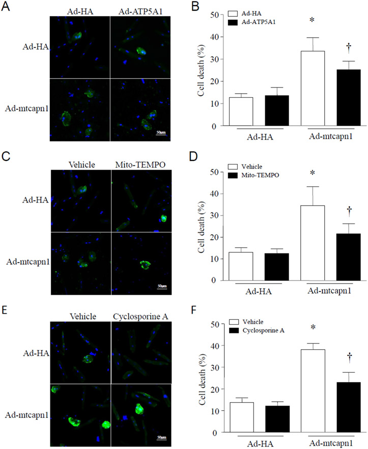
Figure 7. Determination of cell death in cardiomyocytes by annexin V staining.
Adult cardiomyocytes were isolated from mice. After attachment, cardiomyocytes were infected with Ad-mtcapn1 or Ad-HA and then Ad-ATP5A1. After adenoviral infection, the cells were incubated with cyclosporine A, mito-TEMPO or vehicle for 24 hours. Cell death was determined by annexin V staining (green). Nuclei were stained with Hoechst 33342 (blue). (A, C, E) Representative pictures from 3 independent cell cultures. (B, D, F) Percentages of annexin V positive cells. Data are mean ± SD, n = 3. *P < 0.05 versus Ad-HA in the same category and †P < 0.05 versus Ad-mtcapn1 in the same category (two-way ANOVA followed by Newman-Keuls test).

