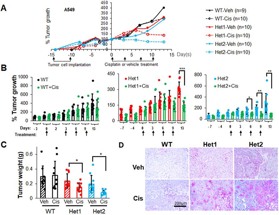
Figure 5: Ceramide kinase inhibition/downregulation sensitizes Mut KRAS NSCLC cells to cisplatin treatment.
(A-D) A549 cells (LUAD histology) with CERK WT (+/+) or Het (+/−) were injected into the flanks of nude mice (106 cells/mice). Mice developing tumors with approximate 100 mm3 volume were treated with cisplatin (3 mg/kg body weight) twice/week for 2 weeks (4 treatments). Tumor size was measured 2-3 times/week (A, B) then mice were euthanized, and tumors were excised for weight measurement (C). Tumor sections were subjected to IHC analysis for ferroptosis marker TFR1 (D). Scale bar = 200 μm. Data in (B,C) are means ± SD; data in (A) are mean; n=9-10 in (A-D). *p<0.05, **p<0.005, ***p<0.0005. Unless otherwise noted by an * or depicted p-value, data are not significant between depicted groups; p>0.05.

