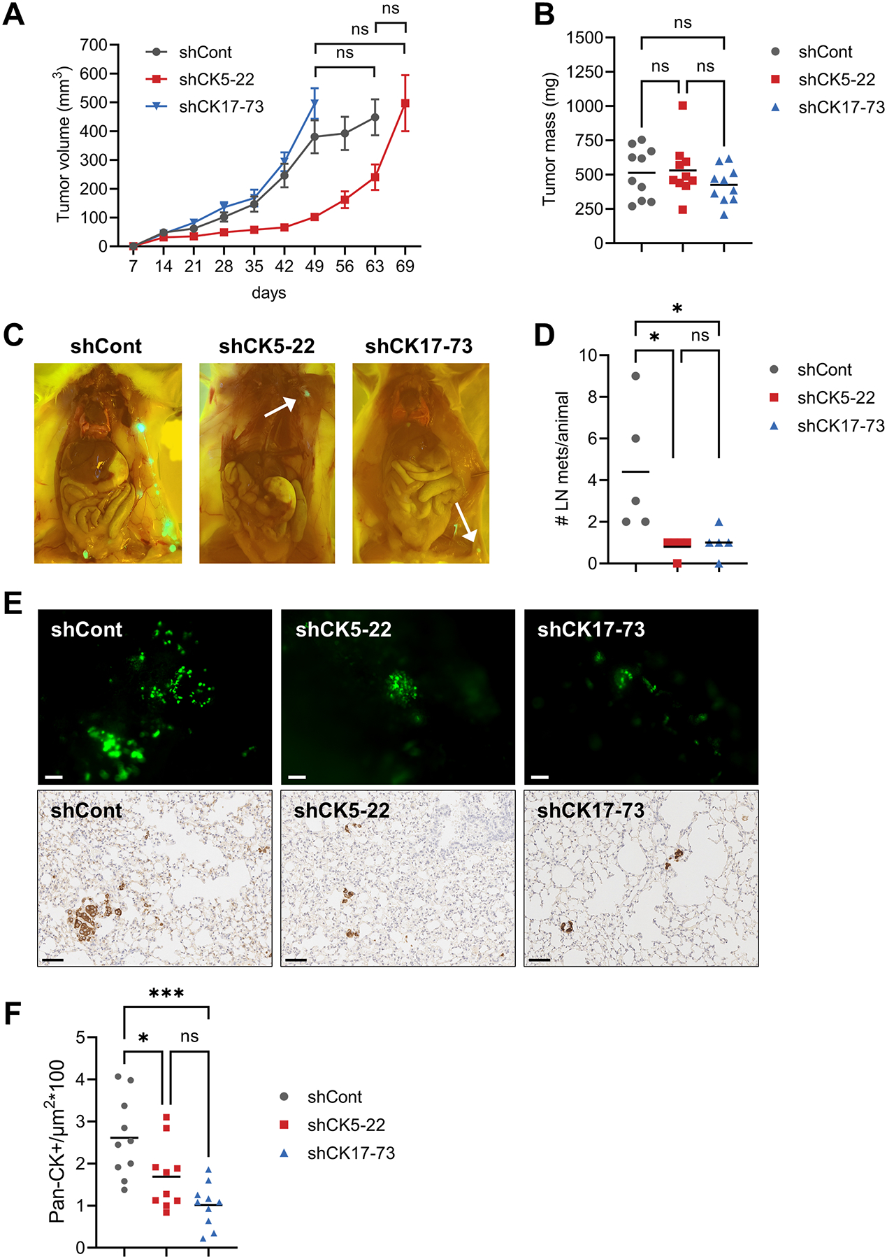
Figure 4. Loss of CK5 and CK17 reduces lymph node and lung metastasis.

A. Growth of GFP-labeled MDA-MB-468 shCont, shCK5-22, and shCK17-73 as xenografts in female NSG mice. Tumors were grown until each group reached an average volume of 500 mm3. Tumor volume over time is depicted. N=10 tumors/group. One-way ANOVA/Tukey test of tumor volumes at the final timepoints is indicated. ns, not significant. B. Final tumor mass for each group. One-way ANOVA/Tukey test. C. Representative images of lymph node metastases at necropsy for each group. D. Number of positive lymph nodes (LN)/animal. One-way ANOVA/Tukey test is indicated. *P<0.05. E. Representative gross images of GFP+ lung metastases in each group (top), scale bars, 100 μm, and representative images of IHC for pan-CK in lung sections for each group (bottom), scale bars, 60 μm. F. Quantitation of lung metastases for each group. Two different sections of lung per animal were stained by IHC with pan-CK antibody and counted as number of strong CK+ cells/μm2 using a trained algorithm on the Aperio digital e-slide analyzer. One-way ANOVA/Tukey was used to determine significance.
