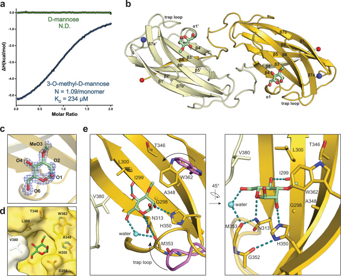
Fig. 2. Molecular structure of Wzt-CBD bound to 3-O-methyl-d-mannose.
a Isotherm plot of Wzt-CBM against 3-O-methyl-d-mannose (blue) and d-mannose (green) Raw data is provided as Source Data. b Overall structure of the Wzt-CBD dimer. Two monomers are colored separately as yellow and vanilla. 3-O-methyl-d-mannose is shown as green sticks. N- and C-terminal ends are shown in blue and red spheres, respectively. c Modeled 3-O-methyl-d-mannose into an unbiased FOFC map. Oxygen atoms are labeled by atom number. Mesh map was contoured at 3σ. d Surface representation of key residues that form the cap sugar-binding pocket. e Two orientations of key interactions between 3-O-methyl-d-mannose and Wzt-CBM. Trap loop and Trp362 from apo Wzt-CBM (PDB ID: 6O14) are colored in violet. The cap-induced movement of the trap loop and Trp362 is indicated with an arrow. Water is represented as a light blue sphere. Hydrogen bonds are shown as dashed lines colored in teal.

