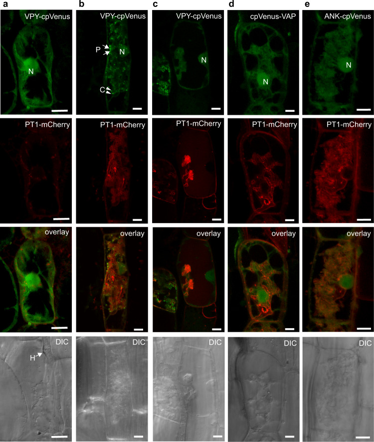
Fig. 1. Subcellular localization of full-length VPY, VAP/MSP, and ankyrin repeat (ANK) domains.
Medicago truncatula roots expressing either VPYpro:VPY-cpVenus (a–c), VPYpro:cpVenus-VAP (d) or VPYpro:ANK-cpVenus (e) (green) and plasma membrane (PM) and periarbuscular membrane (PAM) marker BCP1pro:PT1-mCherry (red) colonized with Rhizophagus irregularis. Images of individual cortical cells prior to and during arbuscule development reveal the subcellular location of VPY (a) before AM fungal entry, (b) formation of fine branches, (c) fully collapsed arbuscule. Fluorescence images are projections of 10 optical sections on the z-axis taken at 0.5 µm intervals. Overlay images display both cpVenus and mCherry channels. Scale bars, 10 µm. H, hyphae; N, nucleus; P, puncta (arrow); C, crescent (arrowhead). DIC, differential interference contrast images of cells. Representative images of two independent transformations, with n = 25 arbuscule-containing cells observed in VPY-cpVenus, n = 43 arbuscule-containing cells in cpVenus-VAP, and n = 45 arbuscule-containing cells in ANK-cpVenus.

