FIGURE 2.

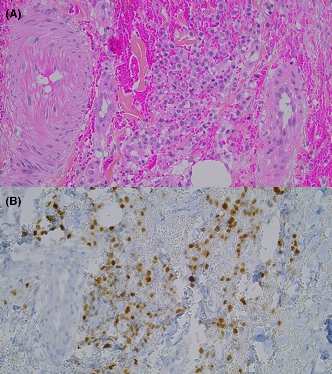
Morphological and immunohistochemical characteristics of the biopsed skin lesion. (A) Focal and perivascular dermal immature plasma cells infiltrate (HPS, 200×). (B) Plasma cells show positivity for MUM1 (IHC, 200×).

Morphological and immunohistochemical characteristics of the biopsed skin lesion. (A) Focal and perivascular dermal immature plasma cells infiltrate (HPS, 200×). (B) Plasma cells show positivity for MUM1 (IHC, 200×).